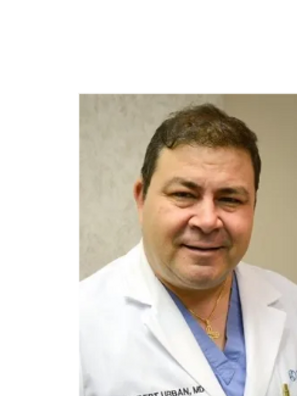
About Dr. Urban
Experienced Medical Professional
Robert C Urban, Jr, M.D. was born in New York. He graduated from Columbia University in 1983 with a Bachelor of Arts. He received his M.D. degree at the Yale University School of Medicine, where he graduated with highest honors, Alpha Omega Alpha, in 1987. Dr. Urban completed his internship in Internal Medicine at the Winthrop University Hospital in Mineola, New York in 1988. He then completed his residency in Ophthalmology at the University of Illinois, Eye and Ear Infirmary in Chicago, Illinois in 1991, then went on to complete his fellowship in glaucoma at Harvard University in 1992.
Dr. Urban is an active member of the American Medical Association, the Association for Research in Vision and Ophthalmology, the American Academy of Ophthalmology, the Tampa Bay Ophthalmologic Society, the American Society for Laser Medicine and Surgery, the Chandler-Grant Society, and the American Glaucoma Society.
Dr. Urban has a keen interest in international ophthalmology. He has served as a visiting ophthalmic surgeon at the request of the governments of: India (Aravind national Eye Hospital), EI Salvador (Rosales National Hospital), and Guatemala (Rodolfo Robles national Eye Hospital). He has presented his work at scientific meetings both nationally and internationally, and his work has been published by the Journal of the American Medical Association, Experimental Eye Research, Investigative Ophthalmology and Visual Science, American Journal of Ophthalmology, International Ophthalmology Clinics, and the Survey of Ophthalmology. He has served as a manuscript referee for Archives of Ophthalmology, American Journal of Ophthalmology, and the Survey of Ophthalmology.
He has served on the faculties of Harvard Medical School, and the University of South Florida. He has served in numerous roles as a consultant, including to the Florida Department of Professional Regulation, and the Ethics Committee of the American Academy of Ophthalmology. He has been named both as one of America's Top Doctors and one of America's Best Doctors.
Compassionate Healthcare
His mission is to provide you with personalized, high-quality eye care. He is dedicated to improving and maintaining your eye health through caring and treating chronic eye diseases while monitoring your eyes for preventive care.
A Personal Approach
His goal is to improve and maintain your overall eye health and to empower you with an understanding of your condition and your treatment plan. Glaucoma Specialist as well as diagnosing other eye conditions such as dry eye, diabetic retinopathy, systemic eye diseases. Call us for more information.

insurances accepted
AETNA COMMERCIAL AND MEDICARE
AMBETTER
BAYCARE PLUS
BLUE CROSS AND BLUE SHIELD (NOT SELECT OR ALIGNMENT)
CHAMPUS
CIGNA
EMBLEM/GHI
ENVOLVE
FARM BUREAU
HUMANA PPO AND HMO COMMERCIAL AND MEDICARE (NO GOLD PLUS)
ICARE
MAILHANDLERS
MEDICARE
MEDICAID
MEDISHARE
MULTIPLAN
MUTUAL OF OMAHA
OPTIMUM AND FREEDOM (MUST BE RELEASED FROM CAPITATON)
PHCS
PREMIER EYE
SIMPLY MEDICARE AND MEDICAID
SOCIAL SECURITY DISABILITY
SUNSHINE MEDICAID AND MEDICARE
TRICARE
UNITED HEALTHCARE ALL PLANS EXCEPT FOR COMMUNITY CARE
WORKMAN'S COMPENSATION (MULTIPLE CARRIERS)
**IF YOU HAVE A MEDICARE SUPPLEMENTAL POLICY NOT LISTED HERE,
CALL TO CONFIRM THAT WE PARTICIPATE WITH IT AS THERE
ARE MULTIPLE SUPPLEMENTAL PLANS**
How We Help
EYE CONDITIONS
Glaucoma
Facts About Glaucoma
Several large studies have shown that eye pressure is a major risk factor for optic nerve damage. In the front of the eye is a space called the anterior chamber. A clear fluid flows continuously in and out of the chamber and nourishes nearby tissues. The fluid leaves the chamber at the open angle where the cornea and iris meet. (See diagram below.) When the fluid reaches the angle, it flows through a spongy meshwork, like a drain, and leaves the eye. In open-angle glaucoma, even though the drainage angle is “open”, the fluid passes too slowly through the meshwork drain. Since the fluid builds up, the pressure inside the eye rises to a level that may damage the optic nerve. When the optic nerve is damaged from increased pressure, open-angle glaucoma-and vision loss—may result. That’s why controlling pressure inside the eye is important.
Dry Eye
Facts About Dry Eye
Dry eye can make it more difficult to perform some activities, such as using a computer or reading for an extended period of time, and it can decrease tolerance for dry environments, such as the air inside an airplane.
Other names for dry eye include dry eye syndrome, keratoconjunctivitis sicca (KCS), dysfunctional tear syndrome, lacrimal keratoconjunctivitis, evaporative tear deficiency, aqueous tear deficiency, and LASIK-induced neurotrophic epitheliopathy (LNE).
Diabetic Retinopathy
Facts About Diabetic Eye Disease
Diabetic retinopathy is the most common diabetic eye disease and a leading cause of blindness in American adults. It is caused by changes in the blood vessels of the retina.
In some people with diabetic retinopathy, blood vessels may swell and leak fluid. In other people, abnormal new blood vessels grow on the surface of the retina. The retina is the light-sensitive tissue at the back of the eye. A healthy retina is necessary for good vision.
If you have diabetic retinopathy, at first you may not notice changes to your vision. But over time, diabetic retinopathy can get worse and cause vision loss. Diabetic retinopathy usually affects both eyes.
Macular Degeneration
Facts About Age-Related Macular Degeneration
In some people, AMD advances so slowly that vision loss does not occur for a long time. In others, the disease progresses faster and may lead to a loss of vision in one or both eyes. As AMD progresses, a blurred area near the center of vision is a common symptom. Over time, the blurred area may grow larger or you may develop blank spots in your central vision. Objects also may not appear to be as bright as they used to be.
AMD by itself does not lead to complete blindness, with no ability to see. However, the loss of central vision in AMD can interfere with simple everyday activities, such as the ability to see faces, drive, read, write, or do close work, such as cooking or fixing things around the house.
Floaters
Facts About Floaters
Sometimes a section of the vitreous pulls the fine fibers away from the retina all at once, rather than gradually, causing many new floaters to appear suddenly. This is called a vitreous detachment, which in most cases is not sight-threatening and requires no treatment.
However, a sudden increase in floaters, possibly accompanied by light flashes or peripheral (side) vision loss, could indicate a retinal detachment. A retinal detachment occurs when any part of the retina, the eye’s light-sensitive tissue, is lifted or pulled from its normal position at the back wall of the eye.
Cataract
Facts About Cataract
What causes cataracts?
The lens lies behind the iris and the pupil. It works much like a camera lens. It focuses light onto the retina at the back of the eye, where an image is recorded. The lens also adjusts the eye's focus, letting us see things clearly both up close and far away. The lens is made of mostly water and protein. The protein is arranged in a precise way that keeps the lens clear and lets light pass through it.
But as we age, some of the protein may clump together and start to cloud a small area of the lens. This is a cataract. Over time, the cataract may grow larger and cloud more of the lens, making it harder to see.
Smoking and diabetes contribute to the development of cataract. Or, it may be that the protein in the lens just changes from the wear and tear it takes over the years.
About Us

Contact Us
The care you deserve.
Please call us for an appointment.
Fax any documents to (727)807-7076
Hours
Monday -Thursday 8:30am to 4:00pm
Friday 8:30am to 12:30pm
Copyright © 2024 urban-eyecare - All Rights Reserved.
Powered by GoDaddy
This website uses cookies.
We use cookies to analyze website traffic and optimize your website experience. By accepting our use of cookies, your data will be aggregated with all other user data.